‘My boy, I wish you to witness an experiment.’ He drew from its case a powerful microscope of French make.
‘What on earth are you going to do, sir?’
The doctor’s brilliant eyes flashed with a mystic light as he replied: ‘Find the fiend who did this crime—and then we will hang him on a gallows so high that all men from the rivers to the ends of the earth shall see and feel and know the might of an unconquerable race of men.’
—Thomas Dixon Jr, The Clansman (1905)
Odile Gaijean was a martyr to her son’s fecklessness. During the 1980s Odile, a resident of Eastern Alsace, had run up huge debts to bail her son out of a financially disastrous stay in Dakar, the capital of Senegal. When he turned up on her doorstep and announced that he was married, his mother was less than impressed. Then his wife turned up – a former prostitute called Fatou Sarre. Domestic bliss it wasn’t. The feud between wife and mother-in-law began with strong words. Soon, though, the women were trading blows. One day Odile, beside herself, picked up a knife. Fatou, not to be outdone, picked up a hammer. Fatou won.
When it was done, she dragged her mother-in-law out of the house and into the barn. Afraid of what might happen to her if her husband found out it was her who killed his mother, Fatou set about destroying the evidence of her crime. In March 1990, on trial for murder, Fatou explained to the court that she did not dare leave Odile’s dying vision intact, for it would surely show her brandishing the hammer. This is why she gouged out her mother-in-law’s eyes.
Three years later, there was a similar case, involving a hapless Cameroonian housebreaker. The press detected a pattern. Was France witnessing a reawakening of ancient ‘Cameroonian legends’ and ‘African beliefs’? Actually, no. On further interrogation, it turned out that Fatou’s ‘beliefs’ – such as they were – dated back to a Bollywood movie she had caught one night in Dakar. It was a whodunnit, based loosely, not on ‘ancient legends’ (Cameroonian or otherwise) but on a much more recent source – European detective fiction.
The short story ‘Claire Lenoir’ caused a minor sensation on its publication in Paris in 1867. Huysmans refers to it in his notorious novel A rebours, and twenty years later, there was still enough milage in the idea that the author, Villiers de l’Isle-Adam, expanded it into a successful novel. Claire Lenoir is the wife of Césaire, an old friend of the story’s narrator, Tribulat Bonhomet. On his way to see them both, Bonhomet befriends Henry Clifton, a young English naval lieutenant who has had himself posted to the South Seas to cure himself of an ill-fated love affair with a married woman who bears more than a passing resemblance to Claire. Later – just before he meets the Lenoirs – Bonhomet stumbles over the following news report:
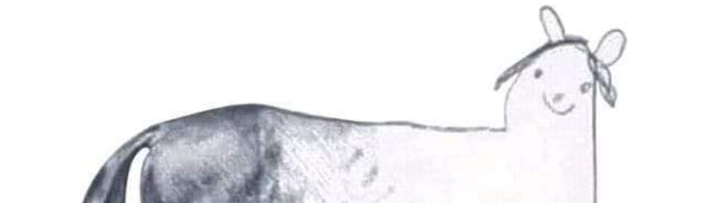
‘L’Académie des Sciences de Paris has stated the authenticity of certain surprising facts. It can be asserted that the animals destined to our nourishment – such as sheep, lambs, horses and cats – conserve in their eyes, after the butcher’s death stroke, the impression of the objects they have seen before they die. It is a photograph of pavements, stalls, gutters, of vague figures, among which one almost always distinguishes that of the man who has slaughtered them…’
It is an ill omen, and sure enough things rapidly take a turn for the worse. His friend Césaire falls desperately ill. Somehow Césaire has discovered his wife’s infidelity; and there on his death-bed, he swears a terrible oath of revenge. A year later, news arrives that Henry Clifton has been brutally murdered by a vampiric assailant. Césaire’s widow Claire takes to her bed, driven out of her mind by persistent nightmares. Bonhomet is with her when she dies and, noticing a blurry image lingering in her eyes, he examines them with the aid of his trusty ophthalmoscope. What he sees,
‘no language, dead or alive… could, under the sun and under the moon, express in its unimaginable horror… I saw the skies, the far-off floods, a great rock, the night and the stars! And upright, on the rock, larger in height than the living, a man… lifted with one hand, towards the abyss, a bloody head, with dripping hair… and, in the severed head, the features, frightfully obscured, of the young man I had known, Sir Henry Clifton, the lost lieutenant!’
It is strange at this remove to imagine the popular curiosity generated by the ophthalmoscope – a device, combining magnifying lenses and a light-source, with which one can view the inside of the living eyeball. The nearest contemporary equivalent would I suppose be the craze, a few years ago, for those ‘magic eye’ posters that conceal three-dimensional figures in a computer-generated pattern of apparently random blobs. They too, as we have seen, have their serious scientific side, and now that the fashion for them is past, they, like the ophthalmoscope, have by and large slipped out of the public gaze, back into the somewhat rarefied world of vision science.
The way de l’Isle-Adam collides the two great scientific crazes of his day – ophthalmology and photography- – is a splendid example of the fantastic at work. The eye is the soul’s camera, and the ophthalmoscope reaches in to spy upon the soul!
If the idea seems trite today, it has only become so through repetition. For de l’Isle-Adam’s readers, ‘Claire Lenoir’ spoke to the bodily anxieties generated by the era’s huge advances in surgery and imaging, as surely and as disconcertingly as the the films of David Cronenberg speak to the bodily anxieties of our own, increasingly anonymous and ‘virtualised’ day. Exposed in this way, the body reveals its affinity with mechanism: the eyes are cameras, and to the well-equipped observer they reveal the secrets of the heart as surely as a daguerrotype reveals in the interior of the sitter’s drawing room. This is a great opportunity – but a risky one, at least in the uncanny world of ‘Claire Lenoir’. At some level, humans are not machines; they are messy, unpredictable, generators of nightmares. You can look, by all means – but you may not like what you find:
‘And Science, the old queen-sovereign with clear eyes, with perhaps too disinterested a logic, with her infamous embrace, sneered in my ear that she was not, she also, more than a lure of the Unknown that spies on us and waits for us—inexorable, implacable!’
Such was the public’s fascination for the new-fangled excitements of forensic science, the fictions that followed ‘Claire Lenoir’ become steadily less like ghost stories, and more like police procedurals. In 1897 Jules Claretie wrote ‘L’Accusateur’, in which the eye of a murdered diplomat, dissected by the police, reveals the blurred image of the face the victim saw as he died. Jules Verne’s Les Frères Kip (1902) sits even more comfortably within the detective genre in its tale of innocent brothers, arrested for a murder they did not commit but released, at the eleventh hour, on the evidence of a retinal photograph. Three years later, retinal photography reveals the identity of a rapist in Thomas Dixon Jr’s jaw-droppingly racist novel The Clansman – the basis for D W Griffith’s film The Birth of a Nation.
Philosophers and scientists could no more resist the analogy between vision and photography than the reading public. In 1859, Charles Darwin’s Origin of Species had explained how, given large enough numbers, time, and a testing environment, living things acquire an efficient, apparently designed form. In acquiring vision, had nature anticipated the very solutions hit upon by those pioneers of photography, Wedgewood, Niépce and Daguerre?
In order for the retina to behave like a camera’s plate, it had to contain some light-sensitive chemical, analogous to the silver nitrate film covering the glass slides on which the earliest photographs – Daguerreotypes – were captured. The presence of a reddish pigment, at least in the retinas of frogs and squid, was established in 1851 by the German anatomist Heinrich Müller (1820–1864). In 1876, a brilliant young compatriot of his, Franz Christian Boll (1849-1879), narrowed the pigment’s source down to one particular group of cells – the ‘rods’. The unusual shape of these cells, coupled with the way they were packed so closely together across the retina, was a clear indication that they possessed some specialised optical function. Were these the cells that saw?
Boll had noticed that when a frog died, the reddish colour of its retinas bleached away, so that after about a minute, the retina appeared colourless. At first he thought this was a consequence of the frog’s death. But then he discovered that even dead frogs retained healthy reddish retinas for up to twenty-four hours, provided they were kept in the dark. If light bleached a pigment stored in the ‘rod’ cells of the frog retina, might that chemical event not convey the impression of light to the frog’s brain? In 1879, Boll’s experiments were cut short: Boll was only thirty when he died, a victim of the tuberculosis that had blighted his otherwise glittering professional life. But he had done enough by 1877 to convince his peers at the Berlin Academy that ‘This change in the outer segments of the rods forms indisputably a part of the process of vision.’ Among Boll’s admirers was Wilhelm ‘Willy’ Kühne (1837-1900). A powerful figure in the field of physiology, Kühne had recently succeeded Hermann von Helmholtz as professor of Physiology at Heidelberg. Kühne took up Boll’s discoveries with ‘fiery zeal’.
Boll had called his pigment ‘visual red’; Kühne reckoned the stuff was more purple than red, and changed the name accordingly. Otherwise, Kühne agreed with Boll – and acknowledged his debt to him – on almost every important point. If the retina was the eye’s ‘photographic plate’, then Boll was surely right, and ‘visual purple’ was the eye’s equivalent of the camera’s silver nitrate. Kuhne hoped to complete Boll’s work by extracting evidence of this process from the retina itself. Working with the simplest equipment in a darkroom, Kühne found a way to maintain retinas long enough that he could study them closely. The dissected retinas remained purple in the dark. Light bleached them, but not all at once. Kühne observed distinct stages in the bleaching process, from purple to orange, to yellow, to buff, until at last the retina became entirely transparent.
Fifty years passed before any new knowledge was added to the meticulous description of visual purple assembled by Kühne over two busy years. Indeed, the pile of unanswerable questions Kühne had amassed concerning the stuff might have altogether obscured his actual achievement, had he not gone to extraordinary and repeated efforts to publicise his central finding: that the eye behaves like a camera.
On November 16, 1880, in the nearby town of Bruchsal, a young man was beheaded by guillotine. The body was taken to Heidelberg, where Kühne, in his capacity as Professor of Physiology, was waiting. In a gloomy room, its few windows screened with red and yellow glass, Kühne dissected the dead boy’s eyes. Ten minutes later, he was able to show colleagues a sharp pattern on the surface of the left retina. This, Kühne told the company, was an optogram: a dying vision, preserved as a pattern in the delicate and unstable visual pigment called visual purple. The trouble was, Kühne’s sketch failed to match any object that may have been visible to the felon as he died.
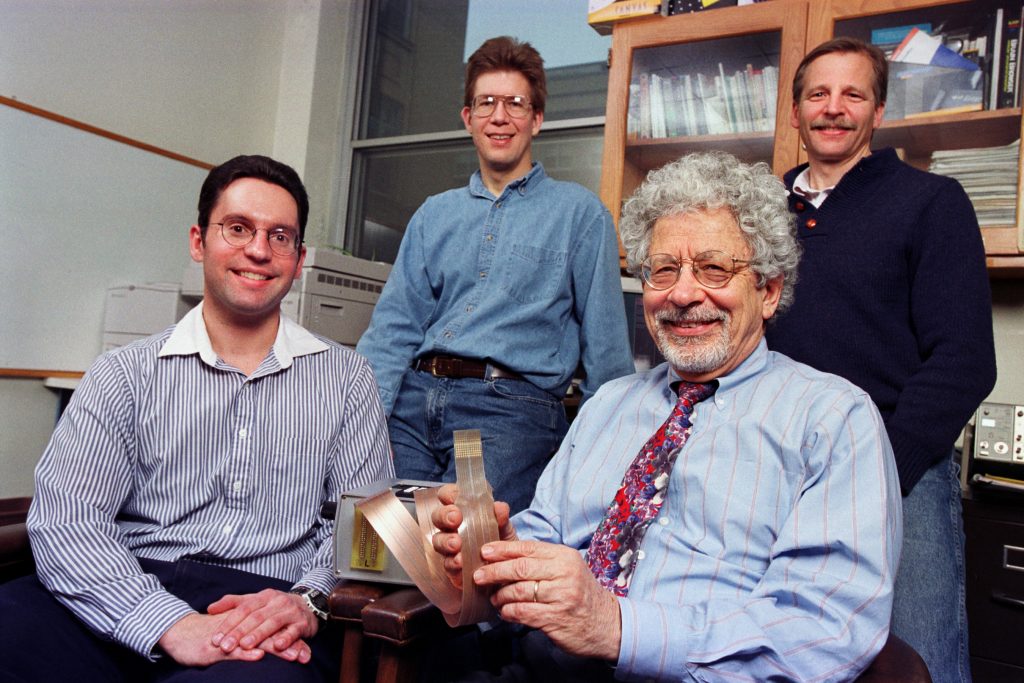
We know now that Kühne had been confronted – and bested – by a curious specialism of the human eye. Its area of focused vision – the fovea – is tiny in comparison to the size of the retina as a whole. Even more puzzling, the fovea is largely rod-free. Kühne had better success recording the optograms of frogs and rabbits. The following account of Kühne’s early and modestly successful ‘optography’ is by the biochemist George Wald – himself a Nobel prize-winner for his studies of visual pigments. (Here, Wald refers to visual purple by its current preferred name: rhodopsin.)
One of Kühne’s early optograms was made as follows. An albino rabbit was fastened with its head facing a barred window. From this position the rabbit could see only a gray and clouded sky. The animal’s head was covered for several minutes with a cloth to adapt its eyes to the dark, that is to let rhodopsin accumulate in its rods. Then the animal was exposed for three minutes to the light. It was immediately decapitated, the eye removed and cut open along the equator, and the rear half of the eyeball containing the retina laid in a solution of alum for fixation. The next day Kühne saw, printed upon the retina in bleached and unaltered rhodopsin, a picture of the window with the clear pattern of its bars. The key word in this passage, of course, is fixation. A living retina has no interest in fixing images. Far from capturing perfect stills, it offers up a constantly changing view. As Kühne himself wrote, ‘the retina behaves not merely like a photographic plate, but like an entire photographic workshop, in which the workman continually renews the plate by laying on new light-sensitive material, while simultaneously erasing the old image.’
It is impossible now to say whether Kühne’s macabre autopsy on the felon from Bruchsal was a rational and necessary extension of his laboratory work, or an aberrant piece of showmanship. Certainly by then his work and the work of his predecessor Franz Boll had been widely popularised in magazines across the globe. In Britain, Fortnightly Nature, Athenaeum and Nineteenth Century ran articles, as did Harper’s Weekly in America. Also, we should not under-estimate the pressure Kühne and other researchers were under to extend the frontiers of forensic science. Since 1857, when the first, probably apocryphal story of optography appeared in Notes and Queries, curiosity about the possibilities of forensic photography regularly found its way into court transcripts. By 1869 the Society of Forensic Medicine in France was so concerned that the courts might start admitting ‘optographs’ as evidence in murder trials, they asked Dr Maxime Vernois to conduct a scientific feasibility study. Vernois set about his task with enthusiasm, ‘violently’ slaughtering seventeen experimental animals ‘under bright lights’. They died in vain: ‘It is impossible to find upon the retina of a victim the portrait of its murderer or the representation of whatever object or physical trait that presented itself to its eyes at the time of death.’
Despite the failures recorded by Vernois and by Kühne, enthusiasm for the idea persisted in spicing up genuine murder investigations. In 1888 Walter Dew – a London policeman famous, years later, for catching the murderer Dr. Hawley Harvey Crippen – witnessed ‘the most gruesome memory of the whole of my Police career’ when he entered 13 Millers Court in Whitechapel, where lay the disordered remains of Mary Kelly, a victim of Jack the Ripper. ‘Several photographs of the eyes were taken by expert photographers with the latest type cameras,’ Dew remembered in his autobiography, in the ‘forlorn hope’ that an image of her killer was retained on the retina. Is Dew’s memory reliable? Perhaps not: but the idea was certainly on everyone’s mind. At the inquest of Annie Chapman, another of Jack’s victims, the jury foreman asked police surgeon Dr George Bagster Phillips, ‘Was any photograph of the eyes of the deceased taken, in case they should retain any impression of the murderer?’ ‘I have no particular opinion upon that point myself,’ Phillips replied. ‘I was asked about it very early in the enquiry and gave my opinion that the operation would be useless, especially in this case.’
There is, as far as I have been able to ascertain, only one, extremely dubious example of a conviction attained by means of an optogram. It is from the Sunday Express and is quoted in Veronique Campion Vincent’s invaluable paper ‘The Tell-Tale Eye’, published in the journal Folklore in 1999. It seems that readers of the edition published on July 4, 1925, thrilled to the tale of Fritz Angerstein, a resident of Limberg in Germany, who killed eight members of his household with a hatchet. The police, seeking a quick confession, told the court that they had taken a photograph of the retinas of Angerstein’s gardener – a picture which revealed ‘a picture of Angerstein with his raised arm gripping a hatchet.’ Hearing this, Angerstein threw in the towel straight away and confessed to the killings.
And the police photograph? Most likely, it was simply a tale, spun to demoralise a gullible defendant. In any event, it was never made public.